Article Text

Abstract
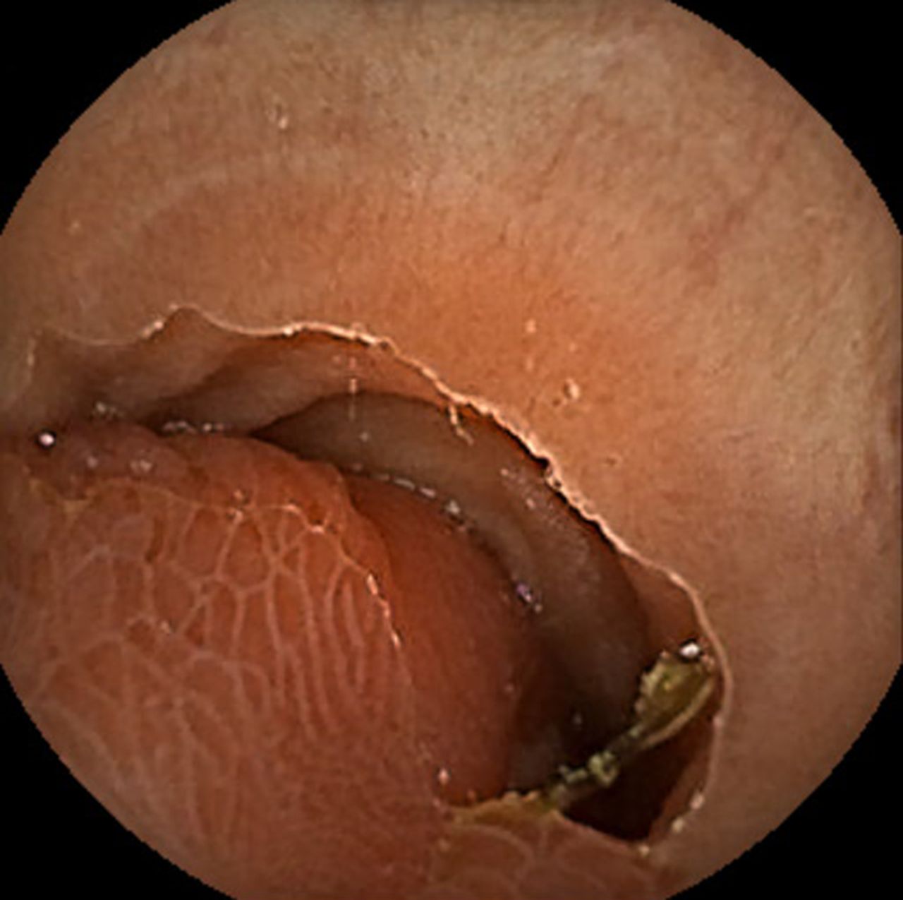
Introduction A 52-year-old woman presented with iron deficiency anaemia and postprandial right lower quadrant pain. Abdominal examination was unremarkable and laboratory results showed mild anaemia (haemoglobin 11.3 g/dL). Upper and lower endoscopies did not reveal any source of bleeding. Video capsule endoscopy was performed which showed a large polypoid lesion in the mid-ileum (figure 1). Abdominal contrast enhanced CT demonstrated a heterogeneously enhancing pedunculated polyp measuring approximately 6 cm (figure 2). Retrograde double-balloon enteroscopy was performed which revealed a large pedunculated polyp with hyperplastic-like mucosa protruding from a large diverticulum located approximately 70 cm proximal to the ileocaecal valve (figure 3A). The stalk appeared to arise from the base of the diverticulum (figure 3B). A technetium-99m pertechnetate scintigraphy revealed no ectopic gastric mucosa.

Video capsule endoscopy shows a large polypoid lesion.
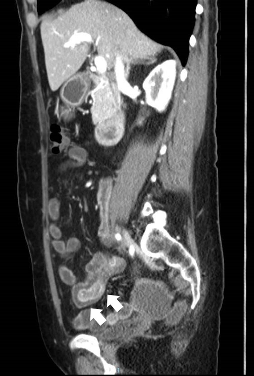
CT shows a heterogeneously enhancing pedunculated polyp (arrow).

Retrograde double-balloon enteroscopy images. (A) Large pedunculated polyp protruding from a large diverticulum. (B) The stalk appears to arise from the base of the diverticulum.
Question What is the diagnosis?
- small bowel
- small bowel disease
- small bowel enteroscopy
- polyp
- diverticular disease
Statistics from Altmetric.com
Footnotes
Contributors YI wrote the paper. YI, TG and NEM treated the patient. CR carried out a pathological assessment of the case. YI, CR, TG and NEM gave final approval of the version to be published.
Competing interests None declared.
Provenance and peer review Not commissioned; internally peer reviewed.