Article Text

Abstract
Objective Percutaneous endoscopic gastrostomy (PEG) tube placement is associated with a high risk of cardiorespiratory complications in patients with significant respiratory compromise. This study reports a case series of high-risk patients undergoing PEG placement using a modified technique—nasal unsedated seated PEG (nuPEG) placement.
Design Retrospective review of 67 patients at high risk of complications undergoing PEG placement between September 2012 and December 2016.
Setting UK specialist tertiary centre for clinical nutrition support.
Interventions Patients underwent ‘push’ PEG placement using nasal endoscopy without sedation in a seated position.
Main outcome measures Procedural success and tolerability, short term (within 24 hours), medium term (24 hours to 30 days) complications and survival were recorded.
Results 67 patients underwent 68 nuPEG placements. The majority had motor neuron disease (46/67). One patient developed a lower respiratory tract infection the following day. Two patients experienced accidental displacement of their PEG within 2 weeks. One patient died within 30 days of nuPEG insertion due to reasons unrelated to the procedure. Endoscopic comfort scores of 1 or 2 (98.0%) indicated good tolerance. A failure rate of 10.5% was attributed to intrathoracic displacement of the stomach, almost certainly due to the advanced stage of the neurological disease and associated diaphragmatic weakness.
Conclusions Our experience with the nuPEG technique suggests that it is safe and well tolerated in high-risk patients. As a result, it has now entirely supplanted radiologically inserted gastrostomy insertion in our institution and we recommend it as the method of choice for gastrostomy tube insertion in such patients.
- artificial nutrition support
- enteral nutrition
- endoscopy
- endoscopic gastrostomy
Statistics from Altmetric.com
Introduction
Progressive neurodegenerative disorders are frequently accompanied by dysphagia that limits oral intake and can result in life-threatening aspiration events. Artificial nutrition support may be required to reduce the risk by permitting reduced oral intake while maintaining nutritional status. Clinical practice guidelines recommend the use of gastrostomy feeding tubes in this setting.1 2 However, the insertion of a percutaneous endoscopically placed gastrostomy (PEG) using standard techniques is associated with significant risk in this patient group due to respiratory muscle dysfunction.3 Cardiorespiratory complications of PEG insertion are responsible for the majority of deaths after the procedure, although they may not be recognised as being directly related to it.4 To reduce this risk, other techniques for percutaneous feeding tube placement are frequently used such as radiologically inserted gastrostomy (RIG) and per-oral image-guided gastrostomy (PIG).5
In view of the increased risks of aspiration associated with the use of intravenous sedation and lying supine during PEG insertion, compounded by the inability to clear secretions with neurodegenerative conditions, we have devised a modified means of PEG placement without sedation and seated upright. We call this technique nasal unsedated seated PEG (nuPEG) placement.
Methods
This study is a retrospective review of all nuPEGs performed at a single institution from September 2012 until December 2016. Cases were identified using the hospital’s electronic database. The patients’ clinical notes were consulted to determine complication rates and mortality data.
All nuPEGs were discussed prior to insertion at a weekly ‘Feeding issues’ multidisciplinary team (FIMDT) meeting where all potential PEG placements are reviewed. Indications and plans for postinsertion care were discussed and agreed at this meeting and a date for insertion confirmed with the endoscopy department. On the day of the procedure, endoscopy staff ensured that the patient had fasted for at least 6 hours prior and any antiplatelet or anticoagulant agents had been held as per the latest British Society of Gastroenterology (BSG) guidance. Consent was obtained for the procedure.
The order of the procedure was as follows:
The patient was sat upright, or at 45% (degrees) in the bed, on the trolley or in their personal wheelchair and the position for tube insertion identified on the abdominal wall. No sedation was given.
Both nostrils were anaesthetised with lidocaine spray.
The nasal endoscope (Pentax transnasal EG16-K10 gastroscope) was passed gently down one nostril and then into the stomach. If required, the mouth was used for suction using the Yankauer sucker.
Patients dependent on non-invasive positive pressure ventilation (NIPPV) devices could still have the endoscope passed with the NIPPV mask in place as it was possible to pass the endoscope without breaking the pressure seal.
Following insufflation of the stomach with the transnasal endoscope, a suitable site for PEG insertion was identified by visualising stomach indentation by external palpation and transillumination where possible. Once identified, local anaesthetic was injected into the skin. A 15Fr Freka Pexact, a direct puncture balloon gastrostomy feeding tube, was inserted using the standard gastropexy placement method. Once the PEG was secure, the nasoendoscope was removed.
Tolerability was measured using a standard endoscopic score of 1–5 where a score of 1 indicates ‘no discomfort experienced—patient resting comfortably throughout’ and a score of 5 indicates ‘extreme discomfort experienced frequently during the procedure’. The comfort assessment was recorded by the endoscopy nurse in conjunction with feedback from the patient after the procedure.
All patients were admitted for overnight stay following nuPEG placement for observation and analgesia. This is routine procedure for all types of PEG insertion in our institution.
Technical success was defined as successful insertion of the nuPEG. Early and late adverse events were defined as arising <24 hours and >24 hours from the procedure, respectively.
Results
Patients
Between September 2012 and December 2016, 67 patients (46 males, 21 females; mean age 70.0 years; age range 39–90 years) underwent 68 nuPEG placements. One of the patients underwent nuPEG twice due to displacement of their original nuPEG. Indications for PEG placement were motor neuron disease (MND) in 46 patients, head and neck cancer with severe respiratory comorbidity in 10 patients, pharyngeal pouch with respiratory comorbidity in two patients, lower oesophageal tumour and respiratory comorbidity in one patient and one patient each with cystic fibrosis, multisystem atrophy, stroke with significant respiratory comorbidity, unexplained oropharyngeal dysphagia and respiratory comorbidity, inclusion body myositis, progressive supranuclear palsy and spinal bulbar muscular atrophy. The median length of follow-up was 429 days (range 21–1480 days).
Success rate
Successful placement was achieved in 60 of the 67 patients (89.5%). Out of the seven patients where nuPEG placement was unsuccessful, six had advanced MND where diaphragmatic weakness had caused migration of the stomach into the thoracic cavity. In these patients, no other PEG insertion technique was deemed appropriate and nasogastric tube insertion was recommended. In the remaining patient, nasal intubation failed.
Early complications (<24 hours)
One patient experienced an episode of desaturation around 18 hours following nuPEG insertion that was treated as a lower respiratory tract infection. This patient had been stable throughout nuPEG placement. This patient recovered with antibiotics and oxygen therapy. No other serious complications were recorded.
Late complications (>24 hours)
Two patients accidently displaced their nuPEG within 2 weeks of placement and required repeat endoscopic sitting. A further patient accidentally displaced his tube 1 month after placement and this was replaced at the bedside. One patient experienced leakage around the tube 1 month following PEG insertion which resolved with topical interventions. There were no reports of peristomal infection within 30 days of insertion.
Tolerability
Forty-one of the 60 successful procedures were given a comfort score of 1 (68.3%). This indicated that the patient was resting comfortably throughout the procedure and experienced no discomfort in the majority of instances. Eighteen patients (30%) were given a comfort score of 2 (one or two episodes of mild discomfort, well tolerated). One patient was given a comfort score of 4 (significant discomfort experienced several times during the procedure)—due to poor tolerance of the passage of the nasal endoscope. Aside from discomfort, this particular procedure passed uneventfully with no complications.
Survival
Sixteen patients died within the follow-up period with a median survival of 136 days (range 21–557). One patient died 23 days after nuPEG insertion due to aspiration pneumonia. This was not thought to be related to the nuPEG placement as he had been well in the intervening period and indicative of the high risk of aspiration due to the underlying disease.
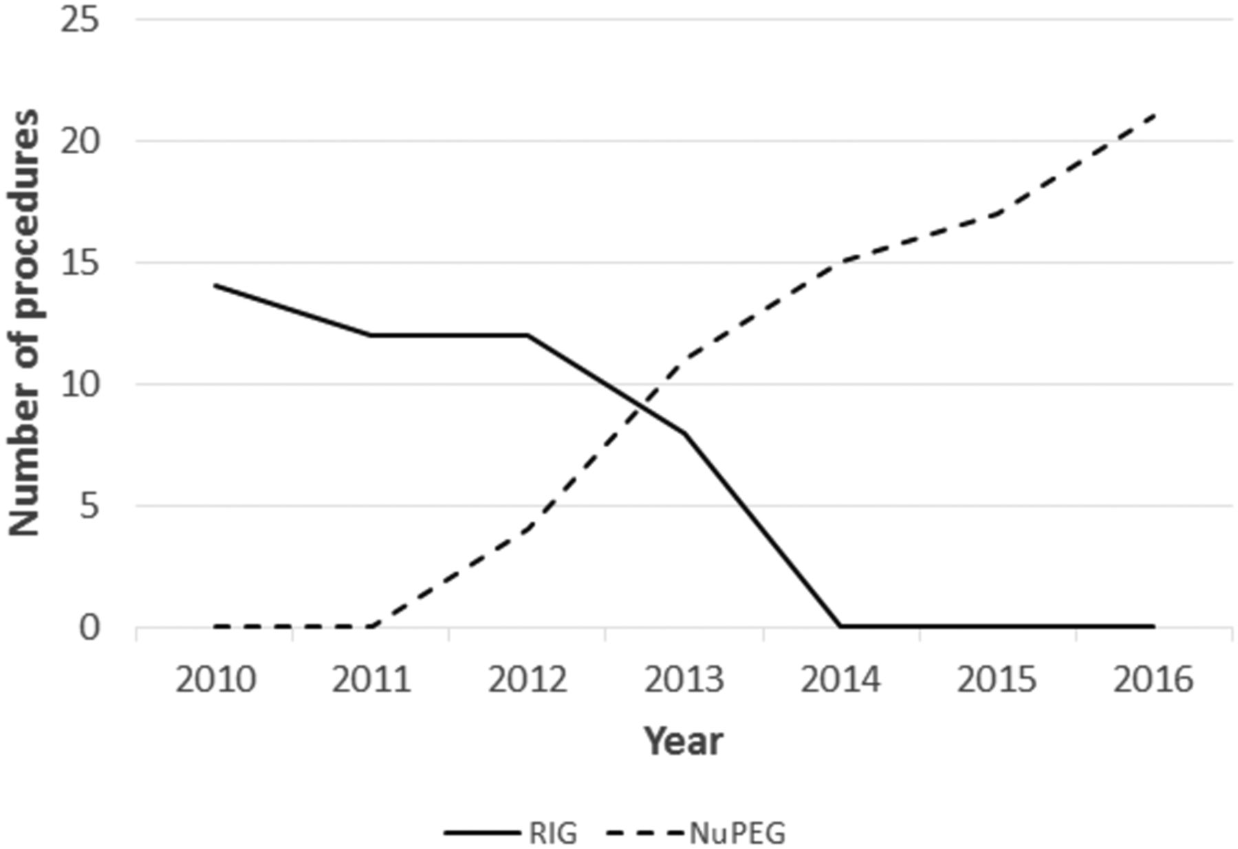
As a result of the success and tolerability of nuPEG placements, this technique has now replaced the use of RIGs in our establishment (figure 1).

The number of radiologically inserted gastrostomies (RIGs) and nasal unsedated seated percutaneous endoscopically guided (nuPEGs) placed in our institution per year over time.
Discussion
Transnasal endoscopy (TNE) has been shown to be better tolerated than standard endoscopy.6–8 This improved tolerability of TNE reduces the need for intravenous sedation making the technique attractive in elderly patients and those with multiple cardiovascular comorbidities. TNE is a simple technique for endoscopists who are competent in conventional endoscopy to learn.9 10 A recent review of the value of TNE noted the lack of studies evaluating its use for PEG placement.11
Previous case series have demonstrated the applicability of unsedated transnasal endoscopy in PEG placement.12–14 Our experience represents the largest such series using TNE in 67 patients over a 4-year period. All patients in our cohort were at high risk of cardiopulmonary complications as a result of significant comorbidity. Favourable outcomes in our patients support the safety of nuPEG placement in such high-risk candidates. Moreover, the only available case series on TNE and PEG insertion describes the use of the ‘pull technique’ using a PEG tube with a collapsible bumper. To our knowledge, this is the only such series to use PEXACT insertion or the ‘push technique’ during TNE which theoretically reduces the potential for peristomal infections as well as nasal trauma.
In patients with MND, RIG or PIG tubes are often used as an alternative to PEG, but there are conflicting reports of their relative safety.15 However, while avoiding the risks of intravenous sedation, lying supine for radiological insertion may not be possible for many patients with severe respiratory compromise and the fixation method and smaller calibre tubes used in RIG placement may increase the risk of obstruction and displacement.16 Thirty-day mortality rates of 3%–8% after PEG placement have been reported in recent series of patients with MND,17–20 although rates as high as 25% have been described21—this compares to 1.5% of the patients in our series.
The nuPEG technique addresses many of the disadvantages of other tube insertion methods in high-risk patients. The procedure is performed with the patient upright and involves no intravenous sedation, therefore, minimising risk of respiratory compromise. Nasal intubation is well tolerated by patients and being awake and able to access the mouth and oropharynx with suction significantly reduces the risk of choking on secretions. Patients feel much more control over the procedure, are able to communicate and interact with the endoscopist and are much more comfortable than with standard PEG insertion. Indeed, the low levels of intravenous sedation that are likely to be given by endoscopists to patients that have been identified as high risk most likely serve only to increase the risks but not to lessen the discomfort of standard PEG placement. Allen et al 18 noted a 10.5% procedural aspiration rate at PEG placement compared with only 1/67 patients in this study following nuPEG insertion. In the PROGAS (prospective gastrostomy) study,19 16% of patients with MND receiving PEGs experienced significant distress during the procedure despite using intravenous sedation. This compares to one patient in this report (1.5%) during NuPEG with the majority experiencing no discomfort at all. Two patients who successfully underwent NuPEG placement had previously failed RIG placement due to panic and inability to lie flat.
The lack of validated patient-reported feedback on the tolerability of the procedure is a potential weakness of this study. However, being unsedated, patients’ feedback (both verbal and non-verbal were unable to speak) was always used by the nurse in the procedure room to record the subjective comfort score. In addition, independent observers (speech and language therapists) who had also witnessed standard PEG placements and RIG placements considered the patient tolerability of nuPEG to be far superior to these two techniques.
For some of the patients who underwent nuPEG placement, it would have been inconceivable to consider any other route of gastrostomy insertion due to the severity of the respiratory compromise. One such patient with cystic fibrosis had previously been suspended from the lung transplant waiting list due to severe undernutrition—RIG insertion proved impossible due to an inability to lie flat and dependence on non-invasive ventilation but following nuPEG placement, he regained sufficient weight to be relisted and transplanted successfully.
A relatively high proportion of patients in this report failed nuPEG insertion (10.5%). Failure rates for PEG insertion are higher in neurodegenerative conditions than for other indications—failure occurred in 6% of patients in PROGAS,19 but 16% of attempted PEG placements in another study.18 We identified intrathoracic stomach displacement as the reason for failure in all but one of our patients and hypothesise that this relates to significant diaphragmatic weakness due to the late stage of disease at which the PEG placement occurred. Whether being seated leads to an increased risk of abdominal compression and gastric displacement is uncertain; however, identification of an appropriate site for PEG placement was not improved by altering the bed conformation to reduce hip flexion.
In addition to low periprocedural complication rates after nuPEG, subsequent complications were also infrequent with no peristomal infections reported on 30-day follow-up. This compares to 16% of patients after PEG placement and 22% after RIG placement in the PROGAS study.19 While this largely relates to the use of Push—rather than Pull—PEGs for the nuPEG technique, one additional benefit of the seated technique is that it is easy to avoid placement of the PEG in skin creases that form when seated which are therefore easily identified.
Due to its excellent tolerability, safety and low periprocedural and postprocedural complications, nuPEG has become the technique of choice for placement of PEGs in high-risk individuals in our institution and as a result no RIGs have been placed since 2013. Furthermore, PEGs have been successfully and safely placed using this technique in patients who were unable to undergo PEG placement by any other route. On the basis of our experience, we recommend the use of nuPEG for all such high-risk patients as the technique of choice.
Significance of this study
What is already known on this topic
Endoscopic gastrostomy tube placement may be contraindicated or associated with a very high risk of complications in patients with significant respiratory compromise.
What this study adds
Gastrostomy tube placement can be safely carried out in high-risk patients using the nasal unsedated seated PEG (nuPEG) technique and is well tolerated.
How might it impact on clinical practice in the foreseeable future
Acceptance of this technique will facilitate gastrostomy tube placement in high-risk patients and result in fewer cardiorespiratory complications. In our institution, it has supplanted radiological placement in this group.
References
Footnotes
Contributors AM and RH collected the data and drafted the article. DM and JMW carried out the procedures with the assistance of OR and AM. SS and NW provided nursing care. JMW devised the technique and revised the draft. All authors read and revised the manuscript.
Competing interests None declared.
Provenance and peer review Not commissioned; externally peer reviewed.