Article Text

Statistics from Altmetric.com
Key messages
Colorectal cancer can occur via more than one molecular pathway. The serrated pathway probably accounts for 20%–30% of colorectal cancer.
Histopathological nomenclature for serrated lesions varies internationally. We suggest the terms hyperplastic polyp (HP), sessile serrated polyp (SSP), and traditional serrated adenoma to describe these lesions.
Colonoscopy is the best detection tool for serrated polyps, but detection rates are variable.
Chromoendoscopy and slower withdrawal time are the only interventions that have been demonstrated to increase serrated lesion detection. High-definition endoscopy and right colon retroflexion may have a role.
All polyps proximal to the recto-sigmoid junction should be removed. A benchmark rate of 4.5% for detection of proximal serrated lesions (HPs plus SSPs proximal to splenic flexure) in screening has been suggested for US-based colonoscopic screening, but implementing a target for serrated lesions in clinical practice is currently impractical.
DNA-based detection significantly augments serrated lesion detection in stool-based screening programmes.
There are limited data to guide surveillance after resection of serrated lesions; however, the logic behind surveillance for serrated lesions is consistent with that for conventional adenomas.
Introduction
A strong evidence base supports colorectal cancer screening. Interruption of the adenoma-carcinoma sequence by endoscopic polypectomy has been considered the key step in preventing the development of colorectal cancer.1 Higher adenoma detection rates (ADR) at colonoscopy have a linear correlation with lower postcolonoscopy colorectal cancer (PCCRC) rates and death from PCCRC.2 However, colonoscopy is not as effective in prevention of colorectal cancer in the right colon as in the left.3–5 Interval cancers are often right-sided and hypermethylated, not consistent with an origin in conventional adenomas.4 Recent molecular approaches to colorectal cancer indicate there are three or more distinct molecular pathways to colorectal cancer, including a pathway arising through serrated lesions.6 ,7
Serrated lesions pose multiple challenges in clinical practice, particularly with regard to detection. Sessile serrated polyps (SSP), the most important subset of the serrated class of lesions, are endoscopically subtle lesions distributed predominantly in the right colon. SSPs are often flat and have minimal discolouration when compared with the background mucosa. These lesions are challenging to detect by any method, including colonoscopy. SSPs are also more challenging to completely resect using standard polypectomy techniques compared to conventional adenomas.8 Finally, there are few observational data to guide development of postpolypectomy surveillance guidelines after resection of serrated lesions.
In this review, we aim to clarify for clinicians the role of serrated lesions in colorectal cancer screening, and the practical issues that flow from our current understanding of their importance.
Terminology
There are two major classes of polyps and flat lesions in the colorectum. The best known class is the conventional adenomas. Conventional adenomas are uniformly dysplastic and, pathologically, the degree of dysplasia is characterised as high or low grade. Conventional adenomas can also be characterised pathologically as tubular or villous. Conventional adenomas are widely understood to be premalignant lesions, with the risk of cancer development increasing in lesions that are larger, or having high-grade dysplasia or villous elements.9
The serrated class of colorectal lesions is distinct from conventional adenomas. WHO recommended that the serrated class has three major subtypes termed as (1) hyperplastic polyp (HP) (2) sessile serrated adenoma/polyp (SSA/SSP) and (3) traditional serrated adenoma (TSA).10 WHO considers the terms SSA and SSP to be synonymous and interchangeable. However, in this review, we will use only the term ‘SSP’, and we recommend that the term ‘SSA’ be abandoned in clinical practice. The term ‘SSA’ had value when first introduced because the word ‘adenoma’ in the term gave importance to the lesion.11 However, the overwhelming majority of SSAs have no dysplastic component. Since all conventional adenomas are dysplastic, the term SSP more accurately distinguishes this generally non-dysplastic lesion from the conventional adenoma. Similarly, the endoscopic surface features of SSP are very similar to HPs, and very distinct from conventional adenomas.12 Finally, the ADR has emerged as the most important quality indicator in the technical performance of colonoscopy.13 Clinicians given a pathology report of ‘SSA’ often believe these lesions should be counted toward the ADR (understandably so when the term SSA includes the word ‘adenoma’), when guidelines clearly indicate the basis and history for why they are not counted toward ADR.13 Thus, based on pathology, endoscopic surface features, and clinical measurements of quality, the term SSA is misleading and confusing, and the term SSP more accurately relates the position of this lesion in the distinct serrated class (box 1).10
Recommended classification of colorectal polyps and flat lesions10
Conventional adenomas
All conventional adenomas can be characterised by their degree of dysplasia (high or low) and tubular vs villous histology
Serrated lesions
Hyperplastic polyps
Sessile serrated polyp
Sessile serrated polyp without cytological dysplasia
Sessile serrated polyp with cytological dysplasia
Traditional serrated adenoma
A distinct and important but small subset of SSPs do contain a region of dysplasia that histologically appears to be a region of conventional adenoma within the SSP. In the past, such lesions were often called ‘mixed hyperplastic-adenomatous polyps.’ The term ‘SSP with cytological dysplasia’ accurately reflects the mixed histology of this lesion which is considered a more advanced lesion in the polyp-cancer sequence than the much more common ‘SSP without cytological dysplasia’.10
The TSA is a rare lesion located primarily in the left colon and rectum.14 ,15 TSA is the only member of the serrated class that is uniformly dysplastic.
Among the two classes of colorectal polyps and flat lesions, only the HP of the serrated class is considered to lack potential for malignancy (table 1). HPs can be subclassified into goblet cell rich type, microvesicular and mucin-poor,10 but this subclassification is not emphasised here because it lacks clinical relevance and is not used by pathologists in clinical practice. However, clinicians may benefit from understanding that the microvesicular subtype is distributed toward the proximal colon relative to the goblet cell rich subtype, and the microvesicular-type accounts for much of the poor agreement among pathologists in differentiating SSP from HP in clinical practice.16 ,17
Clinical features of the major classes of colorectal lesions (conventional adenomas and the serrated class)
The terms ‘polyp’, ‘flat lesion’ and ‘depressed lesion’ are defined in the Paris classification, and are descriptions of endoscopic lesion shapes.18 The use of the term ‘polyp’ in the names of different histological classes of colorectal lesions does not imply that the lesions in that class are all ‘polypoid’ that is, Paris 0-1p (pedunculated) or 0-1s (sessile) in the Paris classification system. For example, conventional adenomas may be polyps, flat lesions, or depressed lesions, and SSPs are either sessile or flat (table 1).
Key molecular aspects
Every cancer is unique from a molecular perspective, but there are three general molecular pathways to CRC.19 The most common is the Chromosomal Instability (CIN) pathway which develops through conventional adenomas and accounts for perhaps 65%–70% of CRCs. Vogelstein developed the concept of chromosomal instabilities leading to progressive accumulation of sporadically acquired mutations in tumour suppressor genes and oncogenes, resulting in development of a conventional adenoma and its transition over many years from low-grade to high-grade dysplasia and then invasive cancer.9
A second major pathway is designated the Lynch pathway and is based in an inherited mutation in one or more of four DNA-mismatch repair genes (MLH1, MSH2, MSH6, PMS2).20 The Lynch pathway, like the CIN pathway, develops through conventional adenomas, but accounts for only about 3% of CRCs. Tumours in Lynch Syndrome consistently demonstrate mutation in short repeating sequences of DNA called microsatellites, and Lynch tumours thus carry a phenotype termed microsatellite instability (MSI). Clinically, MSI appears to be associated with a more rapid transformation of adenoma to cancer, and this observation underlies the recommendation to perform colonoscopy at closer intervals than are needed to intercept cancers passing through the CIN pathway.20
For this review, the third major molecular pathway is of primary interest, since the precursor lesion is serrated. Tumours in this pathway have higher levels of methylation relative to tumours in the other pathways, and the pathway is most often designated the CpG-island methylator phenotype (CIMP) pathway,19 though reference to the ‘serrated pathway’ or ‘hypermethylated pathway’ is also made. DNA-methylation changes cytosin in CpG islands into methylcytosin, which in excess, may inactivate the promoter regions of tumour-suppressor genes (eg, p16, PTEN, E-cadherin, ER, AR, MLH1).21 This event represents epigenetic gene inactivation. About half the tumours in the CIMP pathway have MSI, which results from epigenetic inactivation of MLH1, one of the same mismatch repair genes that when mutated in the germline, results in Lynch Syndrome. Because CIMP pathway tumours are considerably more common than Lynch tumours, CIMP tumours account for about 80% of the CRCs with MSI. The CIMP or ‘serrated’ pathway probably accounts for 20%–30% of colorectal cancer.22–24 Clinicians encountering MSI in tumours may need to differentiate the underlying cause of MSI as CIMP versus Lynch if the affected mismatch repair gene is MLH1. This differentiation can be accomplished by testing the tumour for hypermethylation, or by testing the tumour for mutation in the BRAF oncogene. BRAF mutation is characteristic of CIMP cancers as well as many SSPs and HPs.25
Variable molecular profiles for TSAs have been described, and include the recent finding that TSAs are the sporadic equivalent of hereditary mixed polyposis syndrome, driven by aberrant epithelial GREM1 expression.26
Clinical aspects of the pathology of serrated lesions
Many aspects of the histologic criteria used to classify subtypes of colorectal polyps by pathologists lack validation, and are also subject to substantial interobserver variation among pathologists even when those pathologists are using identical diagnostic criteria. These limitations apply to dysplasia grade and the presence of villous elements in conventional adenomas.27 Clinicians should understand that similar limitations affect the serrated class of colorectal lesions.
First, the agreement between pathologists is low to moderate when differentiating HP (figure 1A) from SSP.16 ,17 The main histologic criteria for diagnosis of SSP are listed in box 2.28 The clinical significance of these diagnostic criteria is not validated. Important features that identify the SSP include lateral growth of crypts at the base, dilation in the lower third of crypts, and hyperserration of the crypt bases, sometimes with branching (figure 1B). Low interobserver variation results when the diagnostic features of SSP are not pronounced or affect only one or a few crypts. To extend the clinical difficulties for clinicians, a multicenter study showed that some pathologists never use the term ‘serrated’ in a pathology report, calling all lesions in the serrated class as HPs.17 Because SSPs are associated with larger size and proximal location compared to HPs, some experts have advocated that lesions larger than 1 cm in size removed from the proximal colon and called HPs by pathologist should be treated as SSPs by clinicians with regard to surveillance.29 This seems particularly reasonable if the institutional pathologists rarely make an SSP determination.
Main histologic diagnostic criteria for sessile serrated polyps (at least 2 out of the 4 criteria for diagnosis in at least 2 crypts not necessarily neighbouring)*
Hyperserration, serration in the lower third of the crypts with and without branching of the crypts
T-shaped and L-shaped crypts above the muscularis mucosae
Inverted crypts (pseudoinvasion) below the muscularis mucosae
Columnar dilation in the lower third
*Modified from Aust et al28 and does not include the ‘side criteria’ in that reference

(A) Hyperplastic polyp with saw-tooth protrusions of the epithelium into the glandular lumen, and eosinophilic cytoplasm. Goblet cells are present but do not reach the basal compartment. The basal zone shows increased proliferative changes with hyperchromatic but not pleomorphic nuclei. (B) Sessile serrated polyp with saw-tooth protrusions of the epithelium into the glandular lumen comparable with hyperplastic polyps but focally with a more complex architecture, and T-shaped or L-shaped glands (black arrow) and dilated glands (yellow arrow) at the base of the lesion just above the muscularis mucosae. Goblet cells reach the base of the lesion in contrast with a hyperplastic polyp. (C) Sessile serrated polyp with cytological dysplasia (low grade). Black rectangle surrounds most of the dysplastic portion. (D) Traditional serrated adenoma showing the typical microacini or microglandular foci and also eosinophilic cytoplasm. Nuclei are hyperchromatic, stratified and palisading, depicting the classical intestinal dysplasia in combination with serration of the tubular glands. This demonstrates a mixture of a distinct subtype of serrated lesion with intestinal dysplasia.
Clinicians should discuss terminology with their pathologists (box 1). We recommend that all SSPs be designated by pathologists as ‘without’ or ‘with’ cytological dysplasia. A focus of cytological dysplasia generally appears to be a region of conventional adenoma within a lesion that is otherwise SSP (figure 1C). Microdissection studies indicate that the dysplastic portion is more likely to demonstrate MSI,30 suggesting that the SSP with cytological dysplasia is an advanced lesion for which an endoscopist should be sure of complete resection. When cytological dysplasia is identified, it may be described as high grade or low grade, but the significance of this designation in SSPs with cytological dysplasia is uncertain. Clinicians should treat any dysplasia in an SSP as a more advanced lesion than an SSP without cytological dysplasia.
The TSA is a rare lesion compared with HP and SSP. Because the TSA is dysplastic and may be villiform histologically (figure 1D), it is likely commonly mistaken for a conventional tubulovillous adenoma, though TSAs have specific features, such as eosinophilic cytoplasm and crypt building.
Detection of serrated lesions during colonsocopy
Endoscopic appearance of serrated lesions
HPs are typically ≤5 mm in size, located in the recto-sigmoid, are hemispherical, often paler than the background mucosa, and usually have either no visible blood vessels or have fine, threadlike branching vessels across their surface12 (figure 2). SSPs are similar in colour and vessel distribution to HPs, but in contrast with HPs, have larger mean size, are distributed toward the proximal colon, have a cloud-like or bossellated surface,31 an irregular surface,31 a mucus cap,32 and indistinct edges31 ,33 (figure 2). Large black pits on the surface of a serrated lesion predict SSP over HP31 (figure 2). Serrated lesions may be recognised by distortion of a fold edge, or disruption of the vascular pattern of the normal colonic mucosa33 (figure 2).
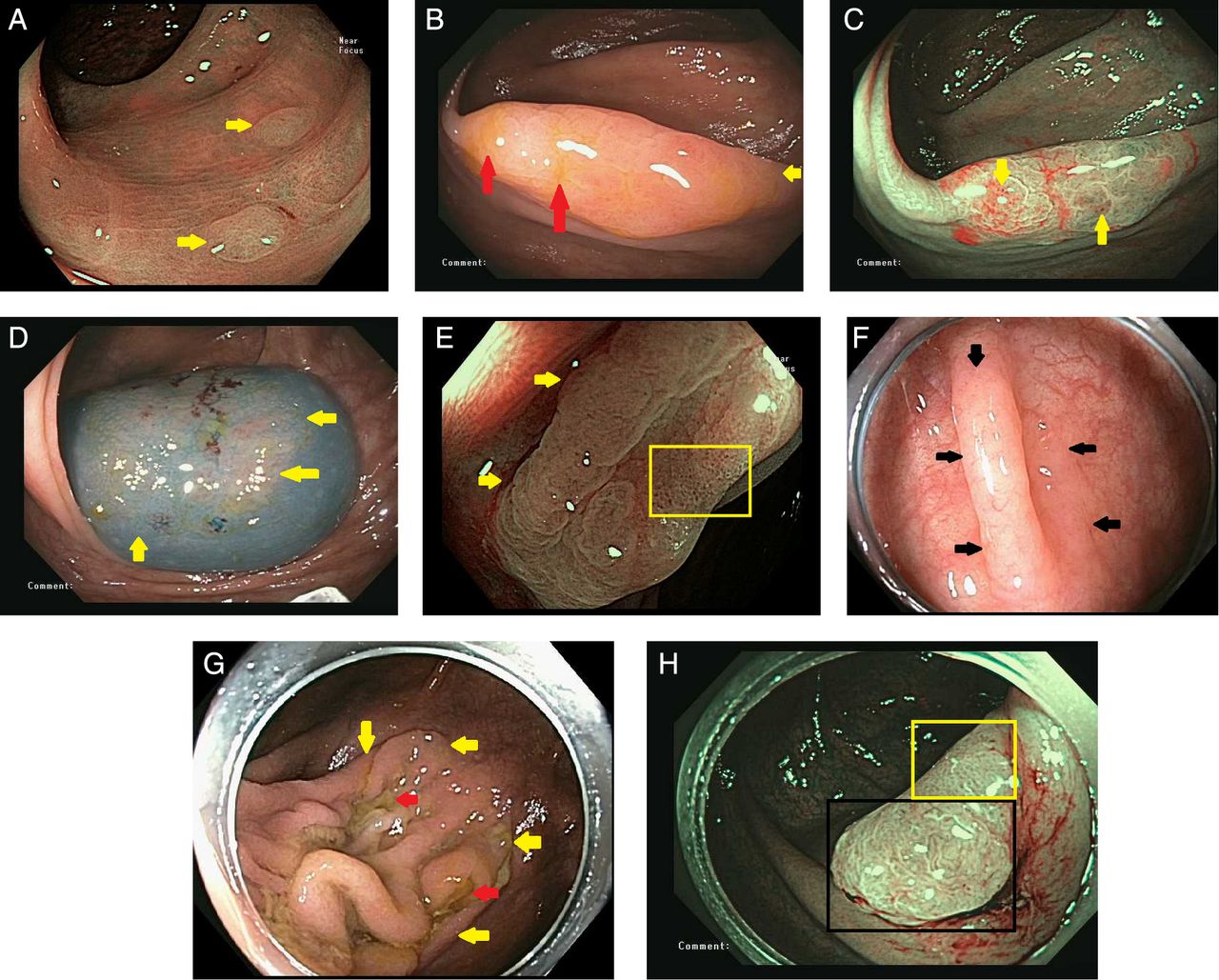
(A) Typical sigmoid colon hyperplastic polyps (arrows) in narrow-band imaging. No blood vessels are visible. The uniform pattern of black dots represent pits. (B) Sessile serrated polyp. Some of the mucus cap remains (orange arrows). One edge is indistinct (yellow arrow). (C) The same lesion seen in (B), now shown in narrow-band imaging. Arrows point to the large dark pits that predict sessile serrated polyp over hyperplastic polyp. The ‘cloud-like’ surface is evident on the left. (D) The lesion seen in 2A and B after submucosal injection with hydroxyethyl starch containing indigo carmine. Arrows show the excellent delineation of the lesion edges by the contrast agent. (E) A sessile serrated polyp in narrow-band imaging. Arrows designate the visible edge. The rectangle encloses a section with numerous large dark pits. The irregular surface of the lesion favours sessile serrated polyp over hyperplastic polyp. (F) A sessile serrated polyp in white light. Arrows delineate the lesion edges. The main clue to the recognition is that the lesion obscures the normal colonic vascular pattern. (G) A very large sessile serrated polyp with the appearance of redundant folds. Yellow arrows mark the lesion edges. The left edge is not visible in the frame. Orange arrows mark areas of residual mucus cap after washing. (H) A sessile serrated polyp with cytological dysplasia. The black rectangle encloses a portion of the lesion with endoscopic features of a conventional adenoma (numerous blood vessels surrounding white pits, which are often tubular, but quite variable in shape). The yellow rectangle encloses a portion of the lesion characterised by uniformly distributed black dots, typical of a serrated lesion.
Colonoscopic prevalence rate of serrated polyps in screening populations
Prevalence rates of proximal colon serrated lesions (HPs plus SSPs) vary between centres17 and between operators within centres,34 ,35 with one US centre reporting a range of 1%–18% among 15 endoscopists performing screening colonoscopy.35 A Dutch screening study with five endoscopists showed a range of 6%–22%.36 The combination of a high detecting colonoscopist and an experienced GI pathologist revealed an SSP prevalence rate of 8.1% in a series of 1910 screening colonoscopies, 0.6% of which had cytological dysplasia.37
Many researchers have chosen to consider all right-sided serrated lesions (HPs plus SSPs) as the outcome measure because of the unreliable differentiation of HP from SSP by pathologists. Prevalence rates of proximal serrated lesions are higher in faecal occult blood test (FOBT)-based colonoscopy programmes, because they are associated with advanced conventional adenomas that are detected by FOBT.38–41 Prevalence rates of proximal serrated lesions vary worldwide from 2.8% to 13% (The Netherlands, Spain, USA, Hong Kong, Korea).17 ,35 ,36 ,38–40 ,42–45 There is also variation between centres within these estimates, with one study of 32 US and German centres reporting a range of 0%–9.8%.17 There may also be differing rates of serrated lesions related to ethnicity, with higher rates reported in Caucasian patients in autopsy studies.46
Serrated polyposis syndrome in screening
Serrated lesions are often multiple, and if in sufficient numbers and size, can meet the criteria for serrated polyposis syndrome (SPS, previously called hyperplastic plyposis syndrome; WHO definition, box 3).47 SPS increases future CRC risk, with Boparai et al48 reporting a 7% risk at 5 years. In FOBT-based screening, the prevalence of SPS may exceed 1:300 (table 235 ,49–52), making this an important syndrome for screening colonoscopists to recognise in these programmes, and again highlighting the association between serrated polyps and advanced adenomas.50 ,51 The rates in colonoscopy-based programmes are approximately 1:2000 (table 2).
WHO definition of serrated polyposis syndrome 201047*
At least five serrated polyps proximal to the sigmoid colon, two of which are greater than 10 mm in diameter
Any number of serrated polyps occurring proximal to the sigmoid colon in an individual who has a first-degree relative with serrated polyposis
More than 20 serrated polyps of any size distributed throughout the colon
*Serrated lesion refers to any combination of hyperplastic polyps and sessile serrated polyps.
Serrated polyposis syndrome prevalence in population-based screening by modality
Serrated polyp miss rates
Few direct measures of serrated lesion miss rates are not available. In 2004, Harrison et al53 found a miss rate for ‘hyperplastic’ polyps of 59% (13/22, 100 patients examined) in the proximal colon. Wide variation in detection17 ,34 ,35 implies large miss rates, as does the increased rate of hypermethylated cancers among postcolonoscopy cancers. Endoscopists using chromoendoscopy find twice the serrated lesions and more proximal serrated lesions as the same endoscopists using white light, indicating that lesions were missed with white light.54–56 Heresbach et al57 determined an overall miss rate for HPs of 31% versus 20% for adenomas. These data indicate that miss rates for serrated lesions may be higher than for conventional adenomas, consistent with their subtle endoscopical appearance.
Technologies and techniques to improve serrated polyp detection
Bowel preparation
Few studies have had detection of serrated lesions as a primary outcome. Quality of bowel preparation does not have the impact on detection of serrated lesions that has been documented for conventional adenomas.58 ,59 In a Dutch screening study, quality of bowel preparation was not associated with lower proximal serrated lesion detection rates, multivariate ORs 0.98 (95% CI 0.92 to 1.05).36 In a US registry-based study, serrated polyp detection rates were similar for optimal (excellent or good) versus fair bowel preparation, with an OR of 0.75 (95% CI 0.31 to 1.80) for poor prep versus optimal prep for proximal serrated polyp detection.60 This effect may result from a thicker mucus cap on serrated lesions with lower quality of preparation.
Withdrawal time
Two studies indicate that longer withdrawal time is important for detection of serrated lesions. In the same Dutch screening cohort, longer withdrawal time had an OR of 1.12 for detection of serrated lesions, identical to that for conventional adenoma detection.36 Analysis of data from the New Hampshire colonoscopy registry reports an incidence rate ratio of 1.77 for each minute beyond 6 min withdrawal time to a maximum at 9 min, slightly higher than that seen for conventional adenomas at 1.50.61
High definition
In a cohort study, detection rates for proximal HPs and for large (≥10 mm) HPs were not different.62 In a Dutch screening cohort, use of high-definition or wide-angle colonoscopes did not improve proximal serrated polyp detection, multivariate ORs 1.07 and 1.30, respectively.36 High definition, compared to standard definition colonoscopy provided only a marginal incremental yield in polyp detection rates of 3.8% (95% CI 1 to 6.7%) in a meta-analysis, suggesting that a major benefit is unlikely.63
Chromoendoscopy
Chromoendoscopy consistently improves adenomatous as well as non-adenomatous polyps detection rates, with the vast majority of the latter being serrated lesions. A summary of four studies performed between 2002 and 2006, before general appreciation of the importance of serrated polyps, suggested an approximate doubling of hyperplastic or non-adenomatous polyp detection from 23% to 45%, and from 9% to 16% when only the proximal colon was considered.56 More recent studies have confirmed this result and effect size in multicentre studies from Germany (46.2% vs 29.5% serrated lesions, rectum excluded, p<0.001)55 and the USA (high-definition colonoscopes; mean non-neoplastic lesions per patient 1.8 vs 1.0, p<0.001).54 The use of chromoendoscopy to increase yield of serrated polyps in the right colon is currently being trialled in an FOBT-positive screening population (CONSCOP Study; ClinicalTrials.gov identifier: NCT01972451).
Narrowed spectrum endoscopy
Narrow-band imaging (NBI) showed promise in a single-centre, single-operator study for detection of serrated lesions in the setting of SPS64; however, a multicentre study from the same group did not confirm this benefit.65 Four different meta-analyses of NBI versus white light, including more than 3000 patients, have failed to show improvements in adenoma or polyp detection rates, suggesting that a benefit for serrated polyps is unlikely.66 Similarly, there is no clear benefit for either flexible spectral imaging colour enhancement (FICE, Fujinon),67–69 or iSCAN (Pentax) with tone enhancement.70–72
Antispasmodics
Hyoscine butyl bromide (Buscopan) increased polyp detection in the right colon, 0.43 versus 0.31, p=0.01 in one randomised controlled trial;73 however, a meta-analysis that included this study found no increase in polyp detection rate overall (OR 1.09 95% CI 0.91 to 1.31).74
Wide angle and proximal colon retroflexion
Proximal colon retroflexion showed a modest increase in proximal serrated lesion detection, although this gain might have been achieved with a repeated examination in the forward view.75 In a tandem study, the Third Eye Retroscope detected 77 versus 58 non-adenomatous polyps, but in the right colon this was only 19 versus 22 non-adenomatous polyps, so better detection of clinically relevant serrated lesions was not shown.76 Other novel devices to reveal more mucosa have been recently reported, such as G-Eye, Full Spectrum Endoscopy and Third Eye Panoramic Device;77 ,78 however, no current studies report data that allow assessment of proximal serrated lesion detection rates.
Bench marks for serrated lesion detection rates
Benchmarks for lesion detection remain a challenge, as a link to important clinical outcomes often requires time to become apparent. Postcolonoscopy colorectal cancer rates are an ideal target for quality improvement but are expensive to measure and fail to detect poor performers early in their experience.79 A Polish screening study validated ADR as a concept associated with CRC prevention.38 ,40 ,80 A larger cohort from the USA found that improvements in ADR above the original recommended thresholds led to additional gains in cancer prevention.2 Initial data suggested that ADR and detection of proximal serrated lesions, those found proximal to the splenic flexure, are highly correlated (R=0.7);35 however, three recent studies found lower correlations (R=0.04–0.43).17 ,43 ,81 Therefore, reaching current ADR targets may not lead to adequate detection of serrations. Kahi and colleagues examined such detections in a cohort of 15 endoscopists for whom the ADR and proximal serrated lesion detection rates were known. They drew a line from the US Multi-Society Task Force ADRs for men (25%) and women (15%) to the regression line and found the equivalent proximal detection rate to be 4.5% for both men and women.35 For programmes based on FOBT, where the higher rate of advanced neoplasia should be associated with higher proximal detection rates, the benchmark may need to be higher.41 One FOBT-based programme reported a rate of 8%.39
While 5% might be a reasonable target for practitioners wanting to check their detection of total proximal colon serrated lesions (HPs plus SSPs), widespread use of this or any proximal colon serrated lesion target would be complicated to institute, since it could be easily corrupted by inclusion of more distal colon serrated lesions (which mostly lack significance).
Endoscopic resection of serrated polyps
Endoscopic resection is recommended for all polyps proximal to the sigmoid colon, all lesions in the recto-sigmoid colon >5 mm in size, and for conventional adenomas in the recto-sigmoid of any size.29 The overwhelming majority of serrated lesions ≤5 mm in the recto-sigmoid are hyperplastic and not SSPs, and recto-sigmoid SSPs ≤5 mm in size with cytological dysplasia are extremely rare. Therefore, serrated lesions ≤5 mm in size located in the recto-sigmoid colon remain the single group of colorectal polyps for which avoidance of resection is appropriate.19 ,29 Identifying this group of lesions and selecting them to be left in situ requires endoscopic estimation of pathology in real time during colonoscopy.12 Experts can exclude conventional adenomas by endoscopic criteria in the recto-sigmoid with >95% accuracy.82
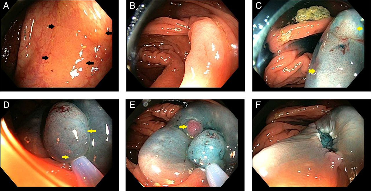
In a recent study of polyps 5 mm–20 mm in size, predictors of incomplete resection were the endoscopists, increasing polyp size and serrated histology.8 The overall rate of incomplete resection of serrated lesions was 32% compared with 7% for conventional adenomas.8 SSPs are characteristically flat or sessile, and have indistinct edges compared with HPs and conventional adenomas.30 ,31 The impact of indistinct edges on incomplete resection is readily understood, but can be overcome by endoscopic mucosal resection employing a contrast agent in the submucosal injection fluid and a high-definition colonoscope.83 This combination provides excellent delineation of the lesion edges, thereby enabling complete resection (figure 3). Inclusion of a margin of normal tissue and the use of snare resection for the entire lesion, with reservation of ablative techniques only for sections that cannot be snared, helps ensure lesion eradication.83 Adherence to these principles is associated with cure rates of endoscopic resection of serrated lesions equal to those for conventional adenomas.83

(A) A sessile serrated polyp with adherent mucus cap. Arrows mark the edges. (B) The same lesion seen in (A) after washing the mucus cap. The edges are less well defined. (C) The lesion after injection with hydroxyethyl starch containing indigo carmine. The edges are now well defined (arrows). (D) A stiff snare is used to facilitate capture of a margin of normal tissue (arrows) around the lesion. (E) The snare is closed very tightly before application of electrocautery. Note the large injection mound. The small red nodule (arrow) is an intramucosal haemorrhage in normal tissue and can be ignored. Larger lesions are resected piecemeal using the same principles. (F) The defect after transection. The blue colour in the submucosa indicates limited thermal injury. The contrast agent and high definition permit visualisation of a tiny nodule of residual serrated tissue (arrow) that requires further snaring or ablation.
Serrated lesions treated with endoscopic mucosal resection techniques are more often resectable en bloc (as opposed to piecemeal) when compared with conventional adenomas of equal size.83
The frequent location of large serrated lesions in the caecum and ascending colon is not a deterrent to their endoscopic resection. There is currently no published evidence that the complication rate of endoscopic resection of serrated lesions is higher than for conventional adenomas; however, concerns have been raised due to the high complication rate seen in a large cohort from pre-2005 describing a subgroup of patients with large sessile lesions resected in the right colon.84 ,85
Resection of serrated lesions by endoscopic mucosal resection is facilitated by use of a stiff snare, which allows the endoscopist to effectively grip the normal mucosa around the lesion and achieve a clear margin (figure 3). Once the snare is closed on tissue, the snare can be squeezed very tightly before application of electrocautery, with less risk of mechanical tissue tearing relative to conventional adenomas. This may be the result of more submucosal fat under serrated lesions compared to conventional adenomas. Some experts recommend tight snare closure before application of cautery to increases current density, speed transection during cautery application, and potentially limit thermal injury to the submucosa.86
Efficacy of other techniques (non-endoscopic) for detection of serrated polyps
When used for screening, flexible sigmoidoscopy generally leads to colonoscopy based on detection of any adenoma, or detection of advanced or multiple adenomas.87 ,88 As such, flexible sigmoidoscopy can detect some proximal cancers and advanced conventional adenomas, with the fraction of advanced proximal lesions detected generally low and dependent on the criteria by which colonoscopy is indicated.87 ,88 One study found that conventional adenomas in the distal colon did not predict advanced serrated neoplasms (SSPs≥1 cm).89 Therefore, available evidence indicates that flexible sigmoidoscopy lacks value for identifying important proximal colon serrated lesions.
Available evidence suggests that detection of serrated lesions by CT colonography is lower than for conventional adenomas.90–92 Studies of CT colonography directly targeting SSPs are not available. Only one trial of capsule colonoscopy directly assessed performance for serrated lesions, and sensitivity was substantially lower than for conventional adenomas.93
Serrated polyps have no or very few blood vessels on the surface,12 and they rarely show haemorrhage pathologically,93 ,94 suggesting they may not be detected by faecal immunochemical testing (FIT). Only one study has directly measured FIT performance for serrated lesions, and the sensitivity of FIT was 5%.95 This sensitivity equalled the false positive rate of FIT, indicating no sensitivity of FIT for serrated lesions.95 Faecal DNA testing includes assays for hypermethylation, and detected 40% of serrated lesions ≥1 cm in size.95 The incremental gain in sensitivity of faecal DNA compared to FIT was much larger for serrated lesions than for conventional adenomas and cancers.95
Surveillance for serrated polyps after colonoscopy
Surveillance for serrated lesions after colonoscopy is controversial because there are no proven benefits, and cost-effectiveness is not established. The guaiac-based FOBT NHS bowel cancer screening programme in England does not endorse surveillance after resection of serrated lesions, citing lack of evidence. Schreiner et al96 reanalysed data collected from 1994 to 1997 in the US Veterans Affairs study group to show that patients who had a proximal serrated lesion alone had an increased risk of future colorectal neoplasia, and those with a serrated lesion and an advanced adenoma had a higher risk of neoplasia and advanced neoplasia at follow-up. Recent data from a Norwegian flexible sigmoidoscopy-based study from an earlier time in which large ‘hyperplastics’ were not considered premalignant suggests that patients with serrated polyps ≥10 mm in size had a future colorectal cancer risk equivalent to having an advanced adenoma, supporting recent guideline recommendations for surveillance of patients with serrated lesions.97 Other circumstantial evidence supports surveillance including: over-representation of the serrated pathway4 in interval cancers, and high rates of residual neoplasia after resection of serrated polyps.8
Given this circumstantial evidence, some but not all editorialists, GI societies, and some international guideline groups have offered guidelines on surveillance (table 3)29 ,19 ,98–100 that are broadly consistent, though the European IARC guidelines make no recommendation.
Cross-tabulation of recommendations for surveillance intervals after detection of serrated lesions
Conclusions and future directions
Colonoscopy is currently not as effective at preventing CRC arising from the serrated pathway compared to CRC arising via the conventional adenoma-carcinoma sequence. However, all other screening methods are less effective than colonoscopy for detection of serrated lesions. With increased understanding of the importance and appearance of serrated lesions, and improved pathological subtyping, detection of serrated lesions is likely to improve. The balance between the risks and costs of resection, and the benefits for CRC prevention remain uncertain.84 High-quality prospective studies are needed to inform surveillance guidelines which are currently based on low or very low-quality evidence. The recognition of a new pathway to CRC outside the traditional adenoma-carcinoma sequence gives researchers and clinicians a chance to further improve CRC prevention by optimising screening to meet this new challenge (box 4).
Key research questions for serrated lesions in colorectal cancer screening
What is the risk of future neoplasia and colorectal cancer after resection of sessile serrated polyps (SSPs)?
Should SSP risks be combined with adenoma risk or additive to adenoma risk for surveillance?
Is surveillance for serrated lesions cost effective?
What chemopreventive measures would reduce the development and growth of serrated lesions and associated cancers?
Is better detection of serrated lesions linked to clinical outcomes?
What tools will increase the detection of serrated lesions at colonoscopy and by other screening methods?
What biomarkers can select patients at high risk of serrated lesions?
How can we teach community-based endoscopists to effectively recognise and safely resect SSPs?
How can we integrate SSP into current paradigms for ‘optical biopsy’ at colonoscopy?
How can we decrease interobserver variability between pathologists assessing SSPs?
How can we link specific molecular changes suggestive of premalignant potential in SSPs to pathological features?
References
Footnotes
Contributors All authors contributed equally to the drafting, critical revision and final approval of this manuscript.
Competing interests DKR receives consulting fees from Olympus Corp, America; Endochoice; and Boston Scientific. He receives research support from Olympus Corp, America.
Provenance and peer review Commissioned; externally peer reviewed.