Article Text

Abstract
Endoscopic ultrasound (EUS) is now firmly established as one of the essential tools for diagnosis in most gastrointestinal MDTs across the UK. However, the ability to provide therapy with EUS has resulted in a significant impact on the management of the patients. These include drainage of peripancreatic collections, EUS-guided endoscopic retrograde cholangiopancreatogram, EUS-guided coeliac plexus blocks, etc. The rapid development of this area in endoscopy is a combination of newer tools and increasing expertise by endosonographers to push the boundaries of intervention with EUS. However, the indications are limited and we are at the start of the learning curve for these high-risk procedures. These therapies should, therefore, be confined to centres with a robust multidisciplinary team, including interventional endoscopists, radiologists and surgeons.
- ENDOSCOPIC ULTRASONOGRAPHY
- PANCREATIC DISORDERS
- PANCREATIC PSEUDOCYST
- PANCREATO-BILIARY DISORDERS
- THERAPEUTIC ENDOSCOPY
This is an Open Access article distributed in accordance with the Creative Commons Attribution Non Commercial (CC BY-NC 4.0) license, which permits others to distribute, remix, adapt, build upon this work non-commercially, and license their derivative works on different terms, provided the original work is properly cited and the use is non-commercial. See: http://creativecommons.org/licenses/by-nc/4.0/
Statistics from Altmetric.com
- ENDOSCOPIC ULTRASONOGRAPHY
- PANCREATIC DISORDERS
- PANCREATIC PSEUDOCYST
- PANCREATO-BILIARY DISORDERS
- THERAPEUTIC ENDOSCOPY
Background
Endoscopic ultrasound (EUS) was developed as a diagnostic modality in the early 1980s for diagnosis of various gastrointestinal (GI) benign and malignant conditions. It is widely used to diagnose/stage malignancies and acquire tissue for diagnosis from pancreas, biliary tree, liver and mediastinum. It has now become an integral part of patient management in multidisciplinary teams across the UK. However, in the last decade, it has rapidly gained a role for providing therapy. Close proximity and relatively safe access to GI and retroperitoneal structures provide opportunity for minimally invasive treatment of various conditions which would otherwise have been treated with other forms of intervention, that is, surgery or interventional radiology.
Due to the wide range of applications, the remit of this paper is to summarise the therapeutic applications of EUS and discuss the future of interventional EUS including training.
EUS-guided drainage of intra-abdominal collections
Drainage of peripancreatic fluid collections
Pancreatic fluid collections (PFCs) develop due to injury to the pancreatic duct in acute or chronic pancreatitis. PFCs are divided into acute peri-PFCs, pancreatic pseudocyst, acute necrotic collection and walled-off necrosis (WON). Pseudocysts and WON usually develop after 4 weeks and if the patient is symptomatic, they would benefit from intervention, that is, drainage which could be surgical, percutaneous or endoscopic route.
Historically, PFCs were drained through surgical cyst gastrostomy and percutaneous drainage either under CT or ultrasound guidance. Endoscopy-guided transmural drainage was first reported in 1975.1 It involved identifying PFC bulge in the gastric wall and creating a fistulous tract using the Seldinger technique. A guide wire is then advanced in to the cyst cavity, and one or multiple stents were deployed to secure the position. EUS-guided drainage of pancreatic pseudocyst was first reported in 1992.2 It allowed the endosonographers to visualise the collection without bulge, avoid blood vessels and ensure adequate apposition for deployment of the stent and was, therefore, thought to be safer.
Since 1992, several studies have reported a success rate of 80%–100% with a complication rate of 10% for pancreatic pseudocysts. A randomised control trial reported that EUS-guided drainage of PFC is associated with reduced length of stay, low morbidity and mortality when compared with surgical cyst gastrostomy.3 However, the complication rate rose significantly from 5% to 30% for infected pseudocysts.4
On the other hand, drainage of WON plastic stents is associated with lower rates of collection resolution and higher adverse event rates. This has been traditionally performed with the insertion of double-pigtail stents and subsequent necrosectomies. However, the patients needed more repeat procedures for complete resolution of the necrotic cavity.5 Newer fully covered self-expanding metals stents (FCSEMS) and lumen-apposing metal stents (LAMS) have now been used for the drainage of PFCs (figure 1).
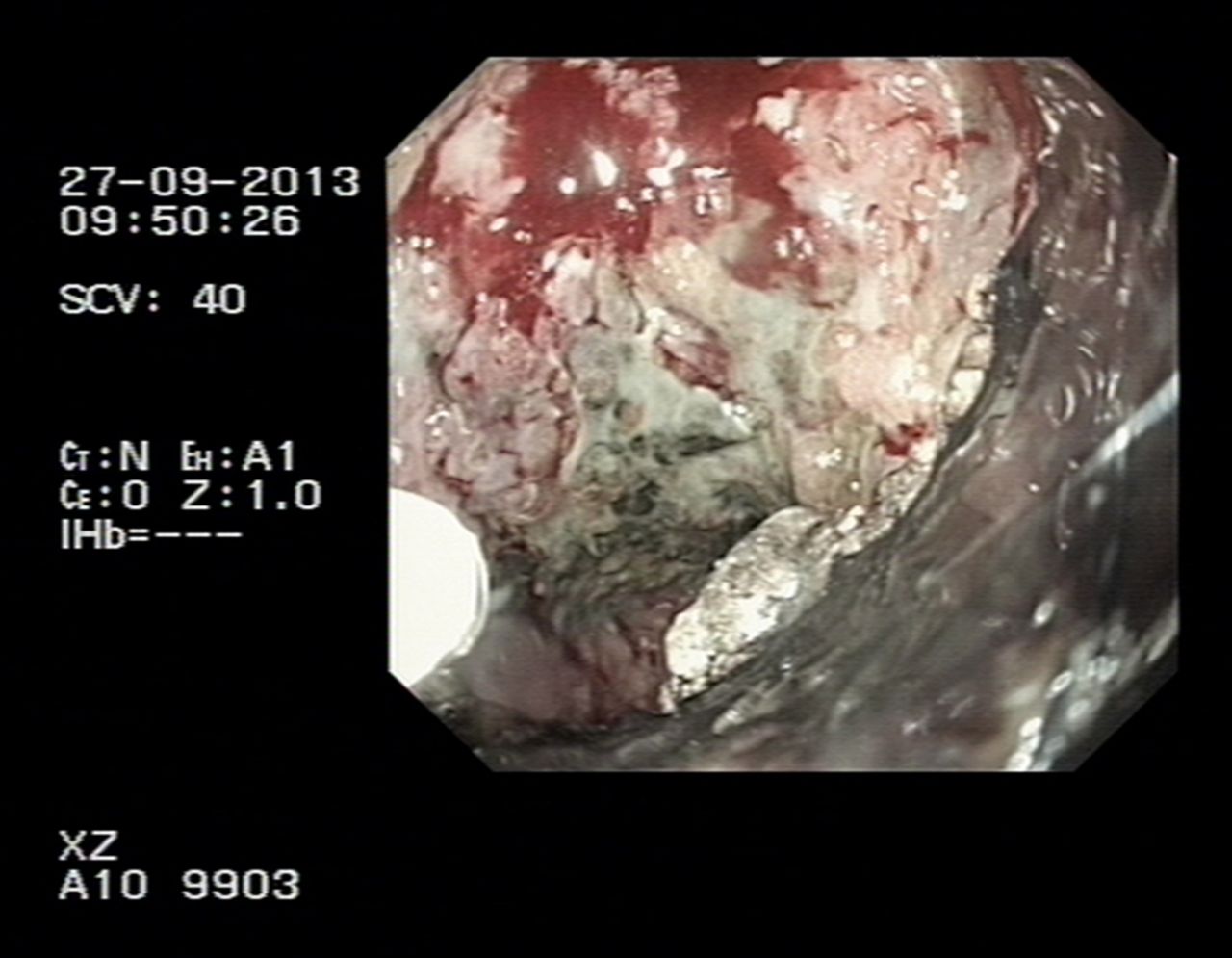
Endoscopic necrosectomy.
Huggett et al6 reported the use of a novel double flanged type of FCSEMS in 19 patients with WON and concluded that the use of this stent is feasible and safe for the drainage of WON. However, stent displacement rates were high, and improvements to the stent design were required before it could be advocated for routine use in WON.
A newer LAMS has been used for the drainage of WON due to its antimigratory properties. A retrospective analysis of 124 patients who had endoscopic drainage of WON by using LAMS concluded that it is safe and highly effective minimally invasive treatment modality for these patients.7
In conclusion, it appears that the minimally invasive approach is associated with an overall decreased mortality rate, fewer major and long-term complications compared with surgery, especially in patients with pseudocysts. However, we need more data on cost-effectiveness and randomised trials to firmly establish the role of metal stents in WON.
Gall bladder drainage
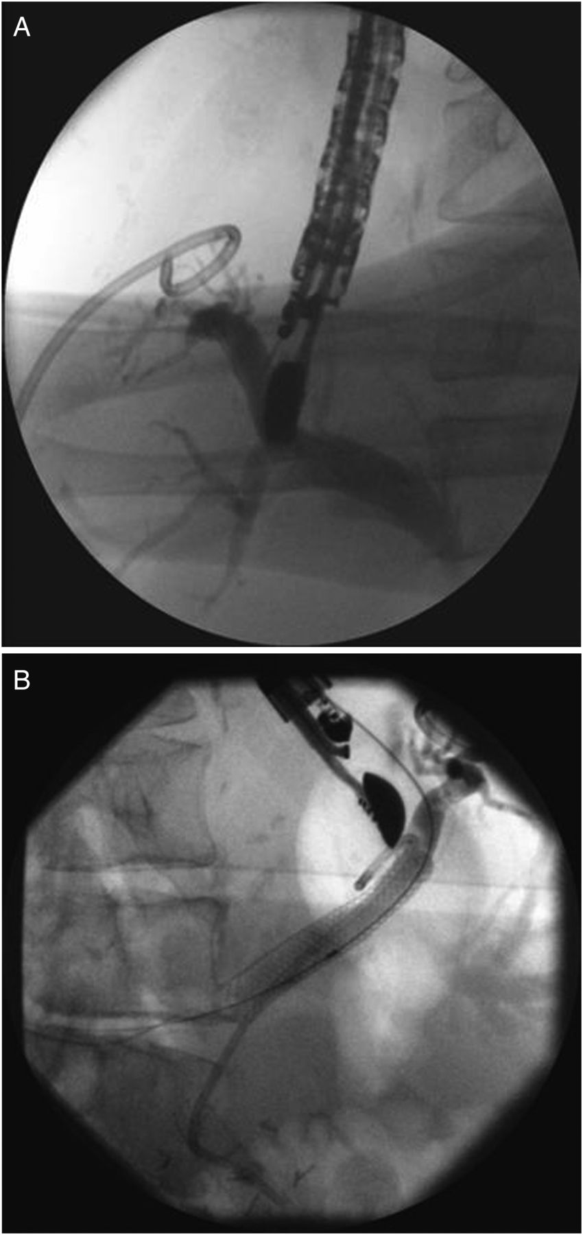
Cholecystectomy is the treatment of choice in patients with acute cholecystitis. However, in certain group of patients, this is not an option due to multiple comorbidities. Traditionally, these patients have been treated with either a permanent percutaneous drain or a cystic duct biliary stent until the patients are fit for surgery. Since it was first reported in 2007, there have been a number of small case series on this form of intervention.8 The recent introduction of LAMS has facilitated ease of insertion (figure 2A, B).9 However, this continues to be a rare indication which should only be considered after careful discussion in a MDM setting.

Endoscopic ultrasound-guided gall bladder drainage—lumen apposing metal stents in GB—endoscopic view and CT view—8 days after insertion.
Other intraabdominal collections
There have also been case reports/series on the drainage of hepatic abscesses, bilomas, subphrenic and pelvic abscesses.10 These are very rare indications for EUS-guided therapy.
EUS-guided biliary drainage
Endoscopic retrograde cholangiopancreatogram (ERCP) is a widely accepted mode of establishing biliary drainage. However, it is not always possible to achieve biliary drainage in 3%–5% of patients because of malignant duodenal obstruction, awkward ampullary position or presence of a diverticulum. Percutaneous transhepatic cholangiogram (PTC) is the established alternative to gain access into the bile duct. A retrospective UK study on 16 363 patients assessing the outcomes of PTC for the palliative relief of malignant jaundice reported that PTC is associated with a high morbidity and mortality (30-day mortality 23%). The emergency readmission rate following the procedure was 20% and 35% experienced serious adverse event.11 EUS-guided biliary drainage seems to be an alternative method in achieving biliary drainage in patients who failed ERCP or who are unfit for radiological intervention.
The three options include:
EUS-guided biliary drainage—transduodenal—for distal biliary strictures.
EUS-guided biliary drainage—transgastric—for proximal biliary strictures, accessed through the left lobe of the liver (figure 3A, B).
EUS-guided rendezvous—for patients who have diverticulum or the ampulla is not clearly visible.

Endoscopic ultrasound (EUS)-guided hepaticogastrostomy—EUS-guided cholangiogram and EUS-guided stent insertion of the intrahepatic ducts.
A recent systematic review of 42 studies involving 1192 patients, assessing the safety and efficacy of EUS-guided biliary drainage, reported a cumulative technical success rate of 95%, functional success rate of 92% and complication rate of 23%. There was no significant difference in the technical success rate and adverse event rate between transduodenal and transgastric approach.12 The common adverse events are bleeding, bile leakage, pneumoperitoneum, stent migration and cholangitis.
EUS-guided biliary drainage provides an alternative to drain biliary tree but despite its success rates, high morbidity such as bile leak, perforation and pneumoperitoneum exists. Hence, these patients should be carefully assessed and should be managed by a multidisciplinary team and should be performed only by experts skilled in both EUS and ERCP.13 In addition, this mode of intervention should not be an alternative in units with a poor ERCP cannulation rates.
EUS-guided coeliac plexus neurolysis/block)
Coeliac plexus neurolysis (CPN) has been done under EUS guidance, and it has advantage over percutaneous approach because the scope can be placed close to the coeliac axis which facilitates coeliac plexus localisation. This helps to place the needle accurately and enhances the spread of injection. A meta-analysis of nine studies involving 221 patients reported that in patients with inoperable pancreatic cancer EUS-guided CPN alleviates pain in 70%–80% of patients at 8 weeks. The pain relief was higher in patients who received injections on both sides of coeliac artery. In patients with chronic pancreatitis, the pain relief was 50%–60% at 8 weeks. In clinical practice, the pain relief is not permanent and it recurs after 8–12 weeks, and therefore it is important to involve the pain team in the early stages to optimise pain before considering CPB.14
EUS-guided treatment of tumours
EUS allows accurate targeting for the delivery of various substances directly into pancreas, liver or subepithelial lesions.
EUS-guided fine-needle injection (FNI) has been reported for the treatment of GI stromal tumours, insulinomas, hepatic metastases, oesophageal cancer, cystic neoplasms of the pancreas and pancreatic adenocarcinoma.
Various biological antitumour agents have been introduced into pancreatic and oesophageal cancers under EUS-guided FNI for control of locally advanced disease. Although long-term results are not well studied, preliminary results suggest that these approaches are generally safe and may prove to be an adjunct or alternative to traditional chemoradiation therapies.
Cyber knife stereotactic radiotherapy has been used to treat lung, mediastinal and pancreatic tumours. It delivers precise directed beams of radiation to the tumour using real-time image guidance. The radiographic markers are placed around the tumour either surgically or using transabdominal ultrasound. However, EUS-guided fiducial placement for locally advanced or recurrent pancreatic cancer seems to be a successful alternative.15 However, this is still in the early stages of being established as a mode of delivering treatment and is only being used in research trials.
EUS in the management of GI bleeding
Upper GI bleeding is a common medical emergency with a mortality of 10%, and therapeutic endoscopy is the main modality of treatment. In clinical practice, it is sometimes difficult to achieve haemostasis, especially in gastric variceal bleeds. If endoscopy fails, then they are often referred for transjugular intrahepatic portosystemic shunt. It is contraindicated in patients with encephalopathy and right heart failure. EUS may be used as an alternative to achieve haemostasis in this group of patients.
The utility of EUS in the setting of GI bleeding has been evaluated in a few small series for specific situations, including refractory bleeding lesions (eg, Dieulafoy's and pancreatic pseudoaneurysm) and oesophageal and gastric varices.16 However, endoscopy should be the mainstay of treatment as the quality of evidence for EUS-guided therapy is low and more research is needed to assess the safety and feasibility.
EUS-guided tissue ablation
There is accumulating data on the EUS-guided ablation of cystic and solid pancreatic tumours but the use of these techniques should be within research protocols until results from larger, prospective clinical trials are available.
Other indications
The other indications for interventional EUS, which are in very preliminary stages, include EUS-guided fine-needle tattoo injection for small tumours, EUS-guided angiography, EUS-guided Botox injection and EUS-guided gastroenterostomy.
Training in therapeutic EUS
Therapeutic EUS is one of the most advanced forms of GI endoscopy. It needs excellent spatial orientation, dexterity, accurate interpretation of sonography images and echoendoscope control. To achieve the above competencies, one should be competent in diagnostic EUS. It needs dedicated focused training in the above modalities. In the UK, with the demands of GI training and acute medicine service provision, it is impossible to deliver adequate supervised training in diagnostic EUS, during their 5 years of training. Although there is now a JAG approved format for diagnostic EUS training, the logistics of providing training in therapeutic EUS has not been addressed.
Conclusion
Therapeutic applications of EUS have exponentially increased over the past decade due to the introduction of newer accessories and echoendoscopes. In addition, the evidence is slowly gathering the role of interventional EUS as the first mode of treatment in certain conditions. This development will continue to grow in the coming years with newer techniques and equipments in the pipeline. However, many of the applications mentioned in this paper are associated with significant risk. It is imperative that these cases are discussed in a multidisciplinary meeting before deciding on the optimum mode of intervention. As training in these procedures is limited, these should be restricted to tertiary centres with experienced endoscopists competent in EUS and ERCP, HPB surgeons and interventional and diagnostic radiologists.
Footnotes
Competing interests None declared.
Provenance and peer review Not commissioned; externally peer reviewed.